
Isolation of Burkholderia cepacia Complex from Pericardial Fluid in a Patient with Lung Adenocarcinoma with Metastasis: A Rare Case Report
Abstract
Burkholderia cepacia complex was isolated from pericardial fluid in a patient with metastatic lung adenocarcinoma, representing a rare opportunistic pericardial infection. Clinical improvement following targeted antimicrobial therapy supported its pathogenic significance and highlights the importance of microbiological evaluation of malignant pericardial effusions.
Article Information
- Received
- Accepted
- Published
Academic Editor: N. R. Rajagopalan, Assistant Professor, Department of Chemistry, St. Joseph’s College of Engineering, Chennai
Checked for plagiarism: Yes
Review by: Single-blind
Copyright © 2026 Mirlekar K, et al.
 This is an open-access article distributed under the terms of the Creative Commons Attribution License, which permits unrestricted use, distribution, and reproduction in any medium, provided the original author and source are credited.
Corresponding author: Mirlekar K, LMMF’s Mai Mangeshkar Hospital —
Competing Interests
The authors have no conflict of interest to declare.
Funding
No specific funding statement was provided by the authors.
Data Availability
No data-availability statement was provided by the authors.
Citation:
Introduction
The Burkholderia cepacia complex comprises environmental non-fermenting Gram-negative bacilli associated with opportunistic infections in immunocompromised hosts 1, 2, 12. Pericardial involvement is extremely rare 3. In tuberculosis-endemic regions, malignant and infectious effusions pose significant diagnostic challenges 4, 13. We report an unusual case of Burkholderia cepacia complex isolated from pericardial fluid in a patient with lung adenocarcinoma with metastasis.
Case Presentation
A 50-year-old female from Moregaon presented with dry cough for two months, fever for one month, and Grade III dyspnoea for three weeks. She had a 25-year history of tobacco (misri) use and occupational exposure to smoke from burning agricultural leaves.
On admission, the patient had cough, cold, body ache, and Grade III dyspnoea requiring oxygen support. Physical examination revealed bilateral crepitations.
Investigations
CT chest demonstrated right-sided pleural effusion, lung atelectasis, mild cardiomegaly, and pericardial effusion.
2D echocardiography showed:
· Normal cardiac chambers
· LVEF 60%
· Mild TR and MR
· Mild-to-moderate pericardial effusion (maximum thickness 2 cm)
· No vegetations or thrombus
Serology
· HIV negative
· HCV negative
· HBsAg positive
· HBV DNA viral load >4000 IU/ml
Tuberculosis Workup
· ZN stain: negative
· GeneXpert: negative
· ADA: 7.5 U/L
Fluid Analysis
· USG-guided pleural tapping: 750 ml
· Pericardial tapping: 300 ml
Cytology and Immunohistochemistry
Pleural fluid cytology revealed Category V malignant cells (IACGRSE 2020).
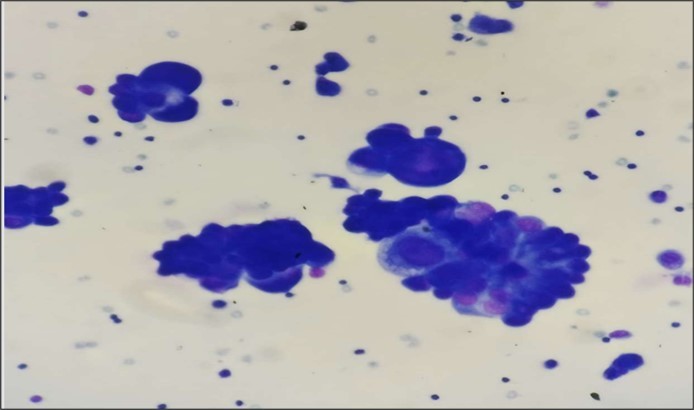
Cytology slides studied from pericardial fluid showed atypical cells with high N: C ratio that are arranged in 3 dimensional balls and glandular pattern. Slides were positive for malignant cells. Confirmation is done by IHC
IHC findings:
· TTF-1 positive
· Napsin A positive
· BerEP4 positive
· PAX8 negative
· GATA3 negative
· Calretinin highlighted mesothelial cells
Diagnosis
Lung adenocarcinoma with metastasis.
PET scan showed FDG-avid consolidative right lung mass with regional nodal involvement, multiple hepatic deposits, and multiple skeletal metastases.
Cytology slides studied from pericardial fluid shows atypical cells with high N: C ratio that are arranged in 3 dimensional balls and glandular pattern. Slides are positive for malignant cells. Confirmation is done by IHC
Microbiology
Sputum culture showed no pathogenic growth.
Pleural fluid cultures demonstrated no microbial growth.
Pericardial fluid culture yielded Burkholderia cepacia complex on Vitek 2. Antimicrobial susceptibility testing was interpreted cautiously, as Clinical and Laboratory Standards Institute (CLSI) 2025 guidelines do not routinely recommend reporting susceptibility for certain antimicrobial agents against this organism 5, 11. However, testing was performed upon clinician request using CLSI 2024 interpretative criteria 6 to guide therapy 11.
The isolate demonstrated:
· Sensitive: trimethoprim–sulfamethoxazole, levofloxacin, meropenem
· Intermediate: minocycline
· Resistant: ceftazidime
The possibility of laboratory contamination was carefully evaluated. The organism was isolated from a normally sterile site obtained via aseptic ultrasound-guided pericardiocentesis. Correlation with clinical findings and subsequent symptomatic improvement following targeted antimicrobial therapy supported its clinical significance rather than incidental contamination.
Pleural fluid cultures remained sterile, suggesting localized pericardial infection rather than disseminated pleuro-pericardial involvement. Isolation of an organism from a sterile body site along with compatible clinical presentation, exclusion of alternative etiologies, and therapeutic response fulfills accepted microbiological criteria for designation as a true pathogen.
Treatment
The patient received:
· Meropenem
· Teicoplanin
Chemotherapy was initiated with:
· Pemetrexed
· Carboplatin
· Granisetron
· Supportive therapy
Following initiation of targeted antimicrobial therapy, the patient showed gradual symptomatic improvement, including reduction in dyspnoea and improved overall clinical status. Oxygen requirement decreased progressively during hospitalization.
Discussion
Burkholderia cepacia complex is an opportunistic non-fermenting Gram-negative bacillus known for intrinsic antimicrobial resistance and survival in moist environmental and healthcare settings 1, 2, 7, 12. Infection predominantly occurs in immunocompromised hosts.
In this case, two states were present. The primary factor was lung adenocarcinoma with metastasis, associated with impaired cellular immunity and malignant effusion formation. The second factor was chronic hepatitis B infection, which may further impair immune responses through chronic inflammation and immune dysregulation 8, 15. The coexistence of these conditions likely predisposed the patient to opportunistic infection.
A key clinical question remains how BCC entered the pericardial space in the absence of cardiac surgery or invasive procedures. Possible mechanisms include hematogenous dissemination from transient bacteremia or contiguous spread from adjacent pulmonary pathology. Agricultural environmental exposure may also have contributed to colonization followed by opportunistic invasion.
An important diagnostic challenge was distinguishing true infection from contamination. Although BCC can exist as an environmental organism, several findings supported pathogenicity in this case:
· Isolation from a sterile anatomical site
· Compatible clinical illness
· Exclusion of tuberculosis and other pathogens
· Sterile pleural fluid cultures
· Clinical improvement after targeted antimicrobial therapy
Together, these factors strongly support true infection rather than laboratory contamination.
Malignant effusions are typically sterile unless secondary infection develops 9, 13. This case highlights the importance of routine microbiological analysis even when malignancy is confirmed. Early identification enabled targeted therapy guided by susceptibility testing performed under clinician request using CLSI 2024 criteria 6.
Tubercular pericarditis was excluded through negative molecular testing and low ADA levels, an essential consideration in endemic regions 4, 10.
Conclusions
Burkholderia cepacia complex may rarely infect the pericardial space in patients with lung adenocarcinoma with metastasis, particularly in the presence of chronic hepatitis B infection. Although malignant pericardial effusions are generally considered sterile, secondary opportunistic infections should be suspected when clinical symptoms persist.
The absence of prior cardiac intervention raises important questions regarding pathogenesis and suggests possible hematogenous or contiguous spread in susceptible hosts. Clinical improvement following targeted antimicrobial therapy further supports the organism’s pathogenic role rather than contamination.
Comprehensive microbiological evaluation of pericardial fluid remains essential for accurate diagnosis and management. Multidisciplinary collaboration between clinicians, microbiologists, and pathologists is crucial for optimal care. Recognition of such rare infections expands clinical understanding and supports evidence-based management of complex oncological patients.14
References
- 1.Mahenthiralingam E, Baldwin A, Dowson. (2008) CG: Burkholderia cepacia complex bacteria: opportunistic pathogens with important natural biology. J Appl Microbiol. 1539-1551.
- 2.Rhodes K A. (2016) Schweizer HP: Antibiotic resistance in Burkholderia species. Drug Resist Updat. 28, 82-90.
- 3.Cunha B A. (2017) Pericardial infections: diagnostic and therapeutic considerations. Infect Dis Clin North Am. 31, 589-601.
- 5. (2025) . Clinical and Laboratory Standards Institute: Performance Standards for Antimicrobial Susceptibility Testing. CLSI M100. 35th ed , Wayne, PA; .
- 6. (2024) . Clinical and Laboratory Standards Institute: Performance Standards for Antimicrobial Susceptibility Testing. CLSI M100. 34th ed , Wayne, PA; .
- 7.Sousa S A, Ramos C G. (2011) Leitão JH: Burkholderia cepacia complex: emerging multihost pathogens equipped with virulence factors. Int J Mol Sci. 12, 6609-6633.
- 8.Rehermann B, Nascimbeni M. (2005) Immunology of hepatitis B virus infection. Nat Rev Immunol. 5, 215-229.
- 9.Maisch B, Seferović P M, Ristić A D. (2015) Guidelines on diagnosis and management of pericardial diseases. Eur. 36, 2921-2964.
- 11. (2025) . Clinical and Laboratory Standards Institute: Performance Standards for Antimicrobial Susceptibility Testing. CLSI supplement M100. 36th ed , Wayne, PA; .
- 12.JEA Zlosnik, Costa P S, Brant R. (2024) Burkholderia cepacia complex infections in immunocompromised hosts: updated epidemiology and therapeutic challenges. Clin Microbiol Rev. 37-00214.
- 13.Imazio M, Gaita F, LeWinter M. (2024) Contemporary management of pericardial diseases: advances in diagnosis and therapy. Eur Heart. 45, 1123-1138.