
Discovery of A New Species of Sap Sucking Plant Mite (Acari: Eriophyoidea) Infesting Medicinal Plant From Sub Himalayan Region of India
Abstract
During general surveys of eriophyoid mites in the Sub-Himalayan region of India, a new species, Diptilomiopus subhimalayansis n. sp. (family Diptilomiopidae), was discovered on the abaxial (undersurface) leaves of Holarrhena pubescens Wall. ex G.Don (Apocynaceae). Because this species is new to science, its morphometric and diagnostic features are fully described and illustrated herein. A brief discussion on the genus Diptilomiopus and the taxonomic relationships of this new species with other related eriophyoid mites are also provided. This new mite species damages its economically important host plant through sap-sucking activity, which causes leaf rusting and brown patches as visible symptoms. This infestation renders the leaves unsuitable for medicinal use. Consequently, taxonomic studies of this pest and a clear understanding of its host plant interactions are essential for developing effective control strategies
Article Information
- Received
- Accepted
- Published
Academic Editor: Devran COSKUN, Siirt veterinary faculty department of pharmacology and toxicology.
Checked for plagiarism: Yes
Review by: Single-blind
Copyright © 2026 Sanjay Sarkar
 This is an open-access article distributed under the terms of the Creative Commons Attribution License, which permits unrestricted use, distribution, and reproduction in any medium, provided the original author and source are credited.
Corresponding author: Sanjay Sarkar, Entomology Research Unit, Department of Zoology, Serampore College under University of Calcutta, Hooghly, (West Bengal) India —
Competing Interests
The authors have declared that no competing interests exist.
Funding
No specific funding statement was provided by the authors.
Data Availability
No data-availability statement was provided by the authors.
Acknowledgements
The author is grateful to Dr Samiran Chakrabarti, Eriophydiologist, Retired Professor, University of Kalyani. Author is obliged to the Principal of Serampore College for providing necessary laboratory facilities during the work.
Citation:
Introduction
The subclass Acari, comprising ticks and mites, represents one of the largest and most biologically diverse groups within the Arachnida. Among these, eriophyoid mites are a highly specialized group of phytophagous (plant-sucking) pests of global agricultural and ecological concern 1, 2, 3, 4. These micro-arthropods cause severe mechanical damage to various plant organs and act as vectors for destructive plant viruses transmitted during feeding 5, 6, 7.
To document the diversity of this fauna, field surveys were conducted across two districts of North Bengal, India—namely Malda and Dakshin Dinajpur, situated within the Sub-Himalayan tract—during October and November 2024. Morphological examination of specimens collected from Holarrhena pubescens Wall. ex G.Don (Apocynaceae) revealed a novel species belonging to the genus Diptilomiopus Nalepa, 1916.
Holarrhena pubescens, commonly known as kurchi or kutaj, is a critically important medicinal tree species indigenous to India. This large deciduous tree is widely distributed across riverine beds throughout the Sub-Himalayan tract, spanning from the Indus River to Assam, as well as adjacent Himalayan valleys. The leaves, stem bark, and seeds of H. pubescens are foundational resources in Ayurvedic medicine 8.
Concurrently, numerous phytochemical compounds extracted from this plant have been cataloged by the Ministry of Ayush (https://www.ayush.gov.in). Recent pharmacological evaluations have validated their efficacy, demonstrating analgesic, antibacterial, antidiarrheal, antidiabetic, antioxidant, antiurolithic, and anti-inflammatory properties 9. Furthermore, contemporary neuropharmacological research highlights its neuroprotective, acetylcholinesterase-inhibitory, and anti-amnesic potential 10.
Taxonomic Diagnosis of the Genus
The genus Diptilomiopus, erected by Nalepa 11, is characterized by a robust, spindle-shaped body and a large gnathosoma with chelicerae abrupt-curved or set perpendicularly relative to leg I. The prodorsal shield is typically wider than long, lacking a frontal lobe, scapular tubercles, and scapular setae, though some species may possess reduced, assetosic scapular tubercles. The legs feature a distinct genu or exhibit genual fusion with the femur; genual setae are invariably absent, leaving only the tarsal setae present. The tarsal empodium is deeply divided. Coxae I may or may not be separated, presenting a less prominent sternal line, and coxal tubercles 1b are absent. The opisthosoma is sub-circular in cross-section, bearing one to three ridges positioned immediately posterior to the prodorsal shield. Opisthosomal setae c2 are missing, though their corresponding tubercles may persist. The epigynium is either smooth or ornamented with longitudinal ribs, lines, granules, or tubercles, while the internal genitalia possess apodemes of normal length.
Diversity and Impact
According to historical catalogs and ongoing faunal checklists of the global Eriophyoidea, over 71 valid species within this genus have been described globally. Of these, 19 species were previously recorded from India 12, 13, 14, 15. However, based on distinct morphological differences distinguishing it from closely related congeners, Diptilomiopus subhimalayansis n. sp. is described herein from the collected specimens.
This discovery increases the known Indian fauna of the genus Diptilomiopus to 20 valid species. This new mite species heavily infests its host plant; its sap-sucking activity induces severe leaf rusting and necrotic brown patches, significantly degrading the quality of the foliage and rendering the leaves unsuitable for phytochemical extraction.
Materials And Methods
Collection and Preservation
Infested plant parts were initially examined in the field using a 30× hand lens. Symptomatic shoots were collected, placed into individual polyethylene bags, labeled with relevant field data, and transported immediately to the laboratory. Duplicate plant specimens were prepared as standard herbaria for botanical identification. To prevent desiccation, fungal degradation, or moisture accumulation within the bags, samples were kept cool, shielded from direct heat, and stored in a refrigerator. When necessary, fresh samples were wrapped in a damp (not wet) paper towel, sealed inside perforated plastic bags to regulate humidity, and processed within a short period.
Slide Preparation and Clearing
The plant samples were examined under a stereomicroscope to confirm mite infestation. Individual mites were isolated from the abaxial leaf surfaces using a fine needle and transferred onto a grooved slide containing Kono’s fluid 16 for clearing. The Kono’s fluid mixture consisted of 100 g chloral hydrate, 10 g glycerin, 50 mL distilled water, and 1 mL concentrated hydrochloric acid (HCl). The slide was subsequently heated on a hot plate at 40 °C until the specimens were sufficiently cleared. Following clearing, the mites were transferred to a bath of Hoyer’s medium and subsequently mounted in the same medium for permanent storage.
Morphological Examination and Morphometry
Morphological attributes were studied using a Leitz Dialux 20 microscope equipped with phase-contrast illumination. Drawings were drafted using a built-in, draw-tube type prism camera lucida. Morphological terminology and abbreviations follow Lindquist 17, while the generic classification system adheres to Amrine et al. 13.
Metric characters were measured at 1000× magnification (10× ocular × 100× oil immersion objective) under phase contrast using a calibrated ocular micrometer. Morphometric data were obtained from 69 specimens, including the holotype, following the standardized protocols recommended by de Lillo et al. 18.
The following morphometric parameters were recorded:
1. Body width: maximum width measured immediately posterior to the rear margin of the prodorsal shield;
2. Body length: distance from the anterior apex of the prodorsal shield to the posterior extremity of the opisthosoma;
3. Prodorsal shield length: distance from the apex of the anterior lobe to the rear margin of the prodorsal shield;
4. Prodorsal shield width: maximum width along the rear margin of the shield;
5. Gnathosoma length: distance from the base of the proximal segment to the apex of the terminal segment;
6. Leg length: distance from the proximal base of the trochanter to the distal tip of the tarsus;
7. Epigynium width: maximum width across the mid-transverse line;
8. Setal length: distance from the basal socket to the apex of the seta.
All measurements are expressed in micrometers (μm) and represent lengths unless stated otherwise. In the taxonomic description, the metric value of the holotype is presented first, followed by the range of the paratypes in parentheses.
Type Depository and Nomenclature
The slide-mounted type specimens were fully labeled and deposited in the Entomology Research Unit (a Biosystematics Research Unit recognized by the Zoological Survey of India as a national depository) at Serampore College, University of Calcutta, 9 William Carey Road, Serampore, West Bengal, India. The new species was registered in ZooBank, and the Life Science Identifier (LSID) was successfully generated (urn:lsid:zoobank.org:pub:6AA8F4DD-54F8-4376-9130-F6D3775BB08B).
Result And Discussion
Based on comprehensive morphometric analysis and a thorough differential diagnosis comparing it with previously described congeners, this mite species is determined to be new to science. Consequently, a detailed taxonomic description is provided herein to formally erect the new species within the genus Diptilomiopus, alongside an account of its host plant interactions. The etymology of the specific epithet adheres strictly to the nomenclature rules established by the International Code of Zoological Nomenclature (ICZN)
Taxonomic description of new species: Diptilomiopus subhimalayansis n. sp. on Holarhena pubescence.
Diptilomiopus subhimalayansis n. sp. is formally described and validated as a phytophagous eriophyoid mite inhabiting Holarrhena pubescens. This description addresses the invalid name Diptilomiopus holarhenansis Sarkar, 2011, which was deemed an unavailable nomen nudum under International Code of Zoological Nomenclature (ICZN) standards, as it was only published in an unregistred Ph.D. thesis. This formal validation provides the necessary, legitimate scientific name for the taxon.
Female (n = 46). Body robust, fusiform, 196.9 (196.9–199.7) long, 84.0 (84.0–87.7) wide; reddish in life. Opisthosoma with four longitudinal ridges and three intervening grooves extending along the anterior two-thirds of the body length (Figure 2 A: LM).
Gnathosoma (Figure 2 A). 23.3 (22.4–23.3) long, abruptly deflected downward; dorsal pedipalp genual seta d 3.7 (3.4–3.7) long.
Prodorsal shield (Figure 2 B: AD). 32.6 (32.6–33.6) long, 59.7 (59.7–60.6) wide, with a distinct anterior shield lobe. Shield design with a complete network of cells; median line incomplete; admedian and submedian lines forming complete cells arranged in four distinct strata over an unequal basal line. Anterior stratum with 12 cells, bisected by the median line; second stratum with 3 cells (one central cell and two subequal, larger lateral cells); third stratum with 3 cells of uniform height but unequal width; fourth stratum with 8 cells of unequal height. Within the fourth tier, the two outermost lateral cells extend anteriorly to touch the first tier of cells. Scapular tubercles and scapular setae sc absent.
Leg I (Figure 2 C). 28.0 (28.0–29.8) long from the base of the trochanter; femur 14.9 (14.9–15.8) long, basiventral femoral seta bv absent; genu fused with the femur; tibia 4.6 (4.6–5.6) long, paraxial tibial seta l' absent; tarsus 7.4 (7.4–8.4) long; paraxial fastigial tarsal seta ft' 33.6 (32.6–33.6) long; antaxial fastigial tarsal seta ft" 31.7 (31.7–32.6) long; paraxial unguinal tarsal seta u' 4.6 (4.6–5.6) long; tarsal solenidion ω 5.6 (5.1–5.6) long, curved downward and knobbed at the apex; tarsal empodium em 7.2 (6.9–7.2) long, deeply divided, each branch 6-rayed.
Leg II (Figure 2 D). 26.1 (25.2–26.1) long from the base of the trochanter; femur 12.1 (12.1–13.0) long, basiventral femoral seta bv absent; genu fused with the femur; tibia 5.6 (4.6–5.6) long, paraxial tibial seta l' absent; tarsus 7.4 (7.4–8.4) long; paraxial fastigial tarsal seta ft' 28.0 (27.4–28.0) long; antaxial fastigial tarsal seta ft" absent; paraxial unguinal tarsal seta u' 4.6 (4.6–5.6) long; tarsal solenidion ω 5.6 (5.1–5.6) long, slightly curved; tarsal empodium em 7.4 (7.1–7.4) long, deeply divided, each branch 6-rayed.
Coxal-genital region (Figure 2 F: CG). Coxae I 22.4 (21.4–22.4) long, contiguous along the midline; coxal surface ornamented with wavy lines near the setal bases; coxal tubercles 1b and setae absent. Coxal tubercles 1a with setae present, situated slightly anterior to a transverse line passing through coxal tubercles 2a; seta 1a 28.0 (26.1–28.0) long. Coxae II 14.9 (14.9–15.8) long, ornamented with two curved lines; seta 2a 43.8 (42.8–43.8) long. External genitalia 20.5 (20.5–21.4) long, 27.0 (27.0–28.0) wide; epigynium ornamented with two parallel wavy lines on each lateral half; seta 3a 6.5 (5.9–6.5) long.
Internal genitalia (Figure 2 G). Internal genitalia characterized by a pair of sub-circular spermathecal pouches and a transverse internal apodeme linked to short spermathecal ducts.
Opisthosoma (Figure 2 A: LM). Dorsum with 68 (66–68) smooth annuli; venter with 85 (83–85) microtubercled annuli. Microtubercles sub-circular, positioned along the anterior margins of the ventral annuli; terminal 9 (9–10) ventral annuli bearing fine microstriations. Setae c2 absent. Setae d 10.2 (10.2–11.2) long, located on ventral annulus 36 (36–37); setae e 7.4 (7.4–8.4) long, located on ventral annulus 54 (54–56); setae f 37.3 (37.3–38.2) long, located on ventral annulus 74 (74–76). Setae h1 absent; caudal setae h2 37.3 (37.3–39.2) long.
MALE. Unknown.
Type Locality: India: West Bengal: Malda, English bazar, Latitude: 24°50′40″ N and Longitude: 87°55′50″ E,
Type material:
Holotype: Female (marked) on slide (no. 1367/57/2024), India: West Bengal: Malda, English bazar, Latitude: 24°50′40″ N and Longitude: 87°55′50″ E, 09.x.2024 from Holarhena pubescence L. (Apocynaceae), Coll. S. Sarkar.
Paratypes: 6 females on slide bearing holotype, 40 females on 5 slides (nos. 1368-1372/57/2024), 14.x.2024, locality of collection same as in holotype.
Figure 1. 1A. Host plant of the Mite: from Holarhena pubescence L. (Apocynaceae); 1B.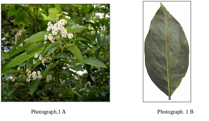
Download figure
Figure 2.Diptilomiopus subhimalayansis n. sp., holotype, 1367/57/2024. : A-Lateral mite (LM); B- Antero-dorsal mite (AD); C- Leg I (L1); D- Leg II (L2); E- Tarsal empodium of LI (em); F- Coxal-genital region (CG); G- Internal genitalia, female. 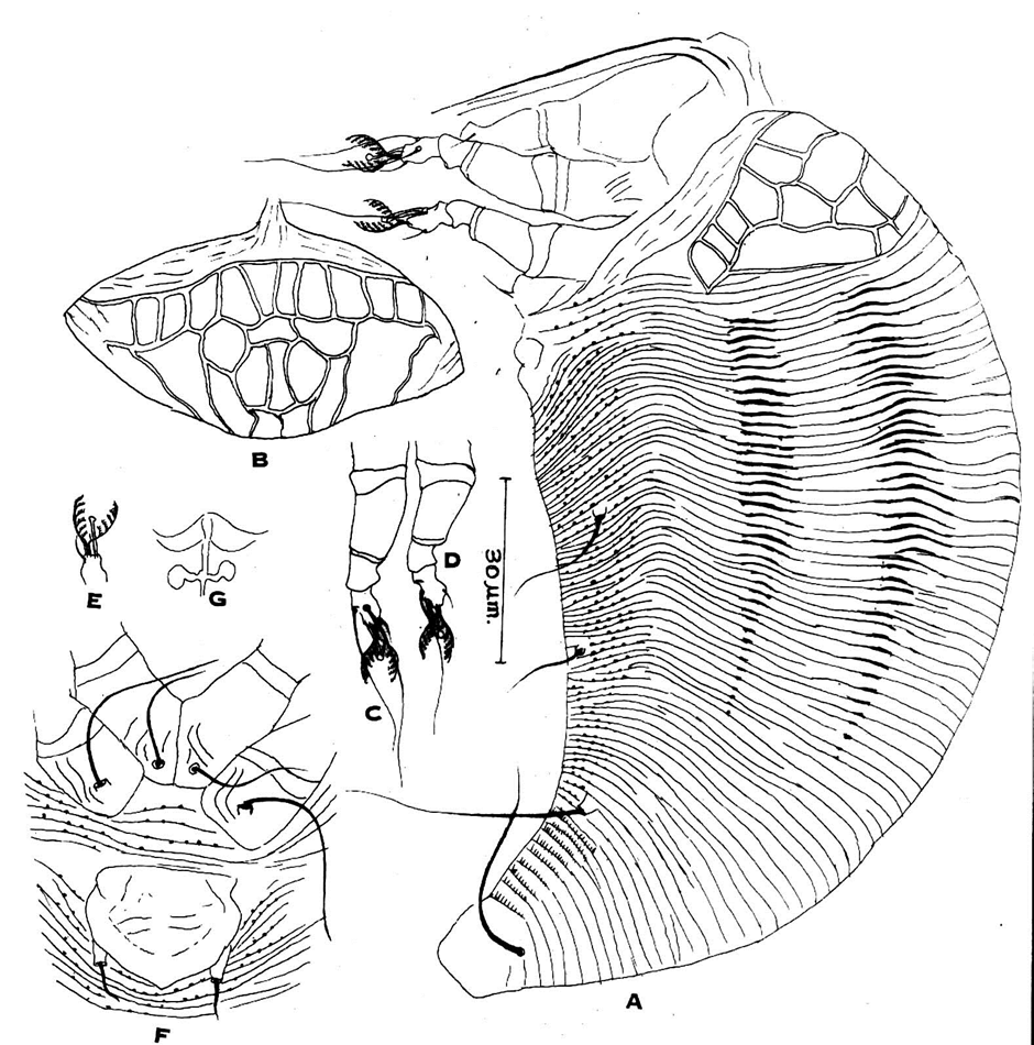
Download figure
Discussion
Differential diagnosis: Globally, the genus Diptilomiopus Nalepa comprises more than 71 valid species, with 19 species previously recorded from India 12, 13, 14, 15. Morphologically, Diptilomiopus subhimalayansis n. sp. belongs to the species group characterized by a distinct cellular network on the prodorsal shield.
The new species can be readily differentiated from D. davisi Keifer, 1969, D. holmesi (Keifer, 1962), and D. jevremovici Keifer, 1960 by the complete absence of scapular tubercles.
Among Indian congeners, Diptilomiopus subhimalayansis n. sp. closely resembles D. ficusis Chakrabarti & Mondal, 1983 in its complex prodorsal shield ornamentation, but it remains distinct from the latter by possessing a 6-rayed tarsal empodium (7-rayed in D. ficusis) and a uniquely distinct epigynium surface ornamentation.
Furthermore, the new species differs from D. trewier Chakrabarti & Mondal, 1983 by the surface textures of both its epigynium and coxae, as well as its 6-rayed tarsal empodium. It can also be easily separated from D. daulatpurensis Sarkar, 2011 by its 6-rayed tarsal empodium
Relation to host: Mites occur as vagrants exclusively on the abaxial (mid-ventral) surfaces of the leaves. Their intensive sap-sucking activity causes prominent leaf rusting and necrotic brown patches (Figure 2 B). Field observations indicated a high localized infestation severity, with symptomatic patches covering a substantial surface area of the foliage. This heavy physical degradation compromises the structural and chemical integrity of the host tissue, thereby rendering the leaves unsuitable for medicinal and phytochemical applications
Etymology:
The specific epithet subhimalayansis is derived from the sub-Himalayan region of India, the broad geographical area encompassing the type locality, and adheres to the nomenclatural rules established by the International Code of Zoological Nomenclature (ICZN).
Conclusion
The present study contributes a novel species of eriophyoid mite to the global documentation of Eriophyoidea diversity, highlighting its significance as an emerging plant pest. Holarrhena pubescens is a critically important medicinal plant indigenous to the sub-Himalayan region of India. The sap-sucking activity of this mite causes severe mechanical and physiological damage to the foliage, which subsequently retards the growth and nutritional uptake of young plants. Consequently, these taxonomic findings provide foundational data that will assist entomologists and plant protection specialists in developing targeted management strategies to safeguard this economically valuable medicinal resource from phytophagous mite infestations
References
- 1.Abdel-Khalek A A. and Momen F M., (2022). Biology and life table parameters of Proprioseiopsis lindquisti on three eriophyid mites (Acari: Phytoseiidae: Eriophyidae). Persian Journal of Acarology 11(1):59-69. https://doi.org/10.22073/pja.v11i1.68574
- 2.Brown, M.S, C.K Blubaugh, and J.H Chong. (2021). Biology and management of eriophyid mites in turfgrass. Journal of Integrated Pest Management, 12(1): 25. http://dx.doi.org/10.1093/jipm/pmab020.
- 4.Vervaet L., R De Vis., P De Clercq. And T Van Leeuwen. (2021). Is the emerging mite pest Aculops lycopersici controllable? Global and genome-based insights in its biology and management. Pest Management Science, 77(6): 2635- 2644. https://doi.org/10.1002/ps.6265.
- 5.Druciarek T., Lewandowski M. and Tzanetakis. (2019). A new, sensitive and efficient method for taxonomic placement in the Eriophyoidea and virus detection in individual eriophyoids. Exp Appl Acarol. 78(2):247–61
- 6.Sarwar M. (2020). Mite (Acari acarina) vectors involved in transmission of plant viruses. Applied Plant Virology 257-273.Elsevier. http://dx.doi.org/10.1016/B978-0-12-818654-1.00020-7
- 7.Stephan D, I Moeller, A Skoracka, F. Ehrig, and E. Maiss. (2008). Eriophyid mite transmission and host range of a Brome streak mosaic virus isolate derived from a full-length cDNA clone. Archives of Virology. 153:181–185. https://doi.org/10.1007/s00705-007-1065-3
- 8.Hamilton AC. (2004). Medicinal plants, Conservation, and Livelihoods. Biodiv. Conserv. 13:1477-1517.
- 9.Ghuge N, Shegar D, Jadhav S. and Nagare V. (2024). International Journal of Pharmaceutical Sciences. 02(12) doi.org/10.5281/zenodo.1441137. Article Id IJPS/240212052
- 10.Bhattacharya S, Saha S, Mukherjee A, Das D, Ghosh S, and Datta S. (2012). Phytochemical analysis and TLC profiling Of Holarrhena pubescens. Pharmacognosy Research.2012;4(2):87-91.
- 11.Nalepa A. (1916). Neue Gallmilben (32. Fortsetzung). Anzeiger der kaiserlichen Akademie der wissenschaften. Mathematisch-naturwissenschaftliche klasse, Wien. 53(22):283-284.
- 12.Amrine J.W. Jr. and Stasny T. A, (1994). Catalog of the Eriophyoidea (Acarina: Prostigmata) of the world. Indira Publishing Houses; Michigan, 798 pp.
- 13.Amrine J.W. Jr., Stasny T.A and Flechtmann C.H.W, (2003). Revised keys to world genera of Eriophyoidea (Acari: Prostigmata). Indira Publishing Houses; Michigan, 244 pp.
- 14.Chakrabarti, S., Sarkar, S. and Pandit, R.K., 2008. Three new species of eriophyid mites (Acari: Eriophyoidea) from Malda district of West Bengal, India. Acarologia, 48(3-4): 203-205.
- 15.Chakrabarti S., Sur S and Sarkar, S. (2019). Two new species of Diptilomiopus Nalepa (Acari: Eriophyoidea) from India. Acarologia 59(3):383-394; DOI 10.24349/acarologia/20194337.
- 16.Jeppson L.R, Keifer H.H. and Baker E.W. (1975). Mites Injurious to EconomicPlants. Univ. Calif. Ber. Los. Arg. London. 614pp.
- 17.Lindquist E.E. (1996). External anatomy and notation of structures. In: Lindquist E.E., Sabelis M.W. & Bruin J. (Eds.), Eriophyoid mites: their biology, natural enemies and control. World Crop Pests, Vol. 6, Elsevier Science Publishers, The Netherlands, pp. 3–31.
- 18.de Lillo E., Craemer C., Amrine J.W. Jr,. and Nuzzaci G. (2010). Recommended procedures and techniques for morphological studies of Eriophyoidea (Acari: Prostigmata). Experimental and Applied Acarology, 51(1–3): 283–307.
- 19.Chakrabarti, S.and Mondal, S. (1983). An account of the genus Diptilomiopus Nalepa (Acarina: Eriophyoidea) from India with description of three new species andkey to the Indian species. Acarologia. 24(3): 299-307.
- 20.Chakrabarti, S., Pandit, R.K and Mandal, S. (2008a). Three new species of eriophyid mites (Acari: Eriophyoidea) from North Bengal, India. Acarologia, 48(3-4): 197-198
- 21.Department of AYUSH. (n.d.). Department of AYUSH. Ministry of Health & Family Welfare, Government of India. [https://www.ayush.gov.in/]. Accessed on 05 March 2025.
- 22.Joel Hallan([email protected]). (2024). A Working Catalogue of the Eriophyoidea of the World. Version 1.0. The Catalogue of the Eriophyidae . Accessed on 05 March 2025.